Pathological findings and distribution of viral antigen in the FMDV-induced hepatorenalopathy in naturally infected cattle calves
218 / 139
Authors
-
 Monalisa Sahoo
Author
Monalisa Sahoo
Author
Keywords:
Apoptosis, calves, FMD, hepatorenal dysfunction, immunohistochemistry, MP-PCR, pathology, serotype-A, serum biochemical analysesAbstract
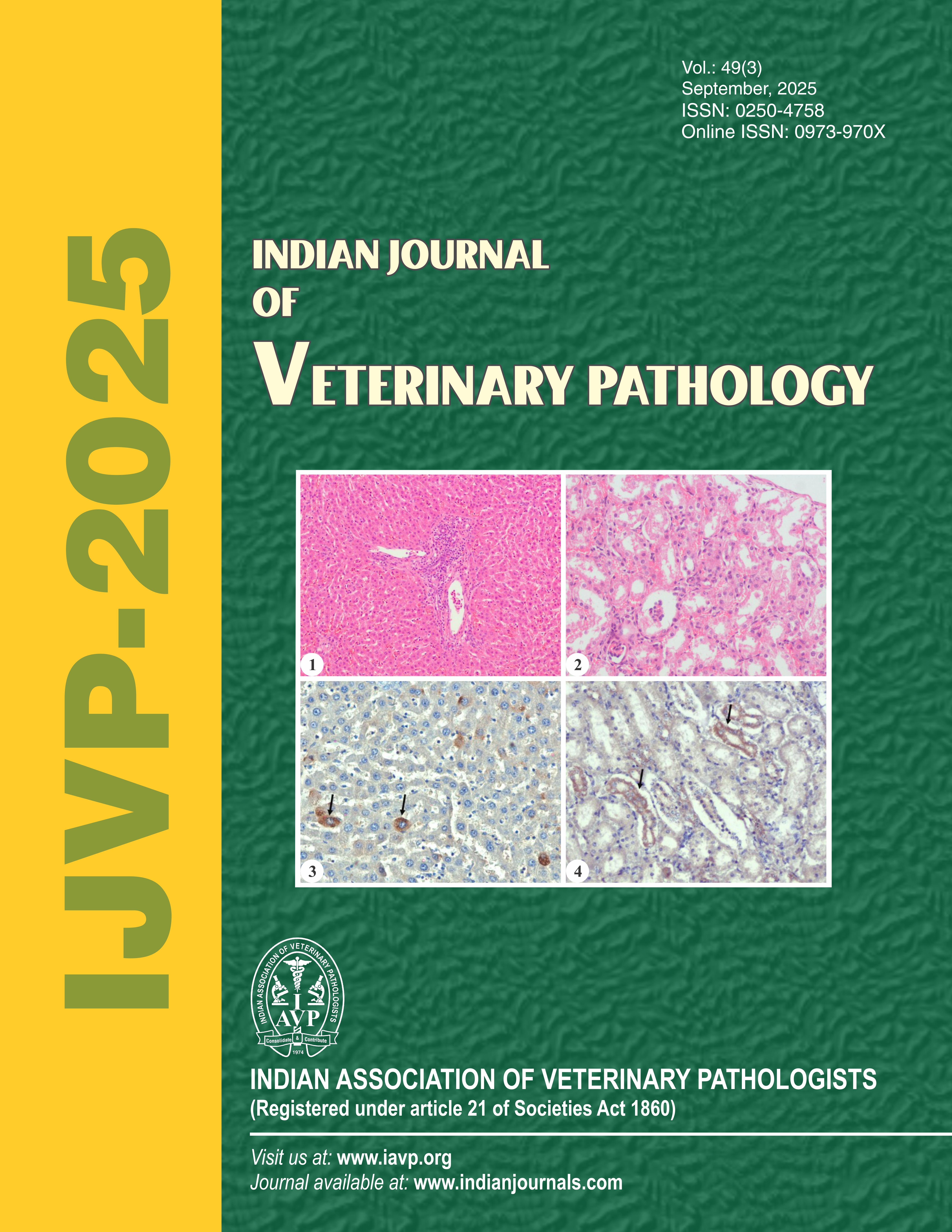
Foot and mouth disease (FMD), the “Risk Group 4” animal pathogen, is causing huge economic loss to the livestock owner due to high morbidity and mortality in young calves. Besides vesicular and cardiac lesions, limited studies are available regarding the FMDV induced hepatorenal dysfunction in the naturally infected cattle. Therefore, the present study was undertaken to investigate the hepatorenalopathy in calves naturally infected with FMDV. The serum from the ailing calves (n=12) were assayed for the estimation of hepatorenal function tests and tumour necrosis factor (TNF)-α. The liver and kidneys from the necropsied calves (n=28) were investigated for pathological, immunohistochemical and molecular investigation along with the virus induced apoptotic changes. The affected calves showed clinical signs of high fever, salivation, vesiculo-ulcerative lesion in the buccal mucosa and skin of hoof cleft. The clinically ailing calves showed elevated serum levels of enzymes in the liver and kidneys (alanine transaminase, aspartate aminotransferase, alkaline phosphatase, blood urea nitrogen, urea, creatinine) and cytokine TNF-α. Post-mortem observations showed the classical lesions of acute necrotizing myocarditis, vesicular/ulceration lesions in the buccal mucosa and clefts of hooves, comparable with the FMD virus infection. In addition to that, variably enlarged livers with rounded borders, centrilobular haemorrhage/necrosis with lobulation, multifocal hepatitis and fibrosis along with congested/hemorrhagic and oedematous kidneys were observed in majority of the calves. Microscopically, the classical lesions of vesicular inflammatory oral lesions, acute necrotizing myocarditis and skeletal muscle necrosis (tongue) were prominent. In addition to that, liver showed prominent multifocal centrilobular necrosis and haemorrhage, surrounded by degenerated fat-laden hepatic cells (like hypoxic changes), multifocal periportal infiltration of mononuclear cells, variably bridging fibrosis and increased activity of Kupffer cells in the sinusoids. The kidneys showed vascular changes such as congestion, oedema and haemorrhages, vasculitis, glomerulitis, interstitial nephritis and tubular degeneration. The presence of viral antigens in the hepatocytes and kidney tubular epithelial cells by immunohistochemistry along with associated elevated serum enzymes support the role of FMDV, causing hepatorenal injury/dysfunction. The TdT-mediated dUTP nick-end labeling (TUNEL) assay confirmed that the death of the hepatocytes and tubular epithelial cells is due to apoptosis. The viral genome in both the liver and kidneys was confirmed to be type A FMDV in multiplex-PCR assay. These results provide insights into novel tissue tropisms in the liver and kidneys of young calves with natural FMDV infections. Therefore, therapeutic intervention should be directed to boost the liver and kidneys for better management of the infected cases.
Downloads
Downloads
Submitted
Published
Issue
Section
License
Copyright (c) 2025 Indian Journal of Veterinary Pathology

This work is licensed under a Creative Commons Attribution-NonCommercial-ShareAlike 4.0 International License.
How to Cite


