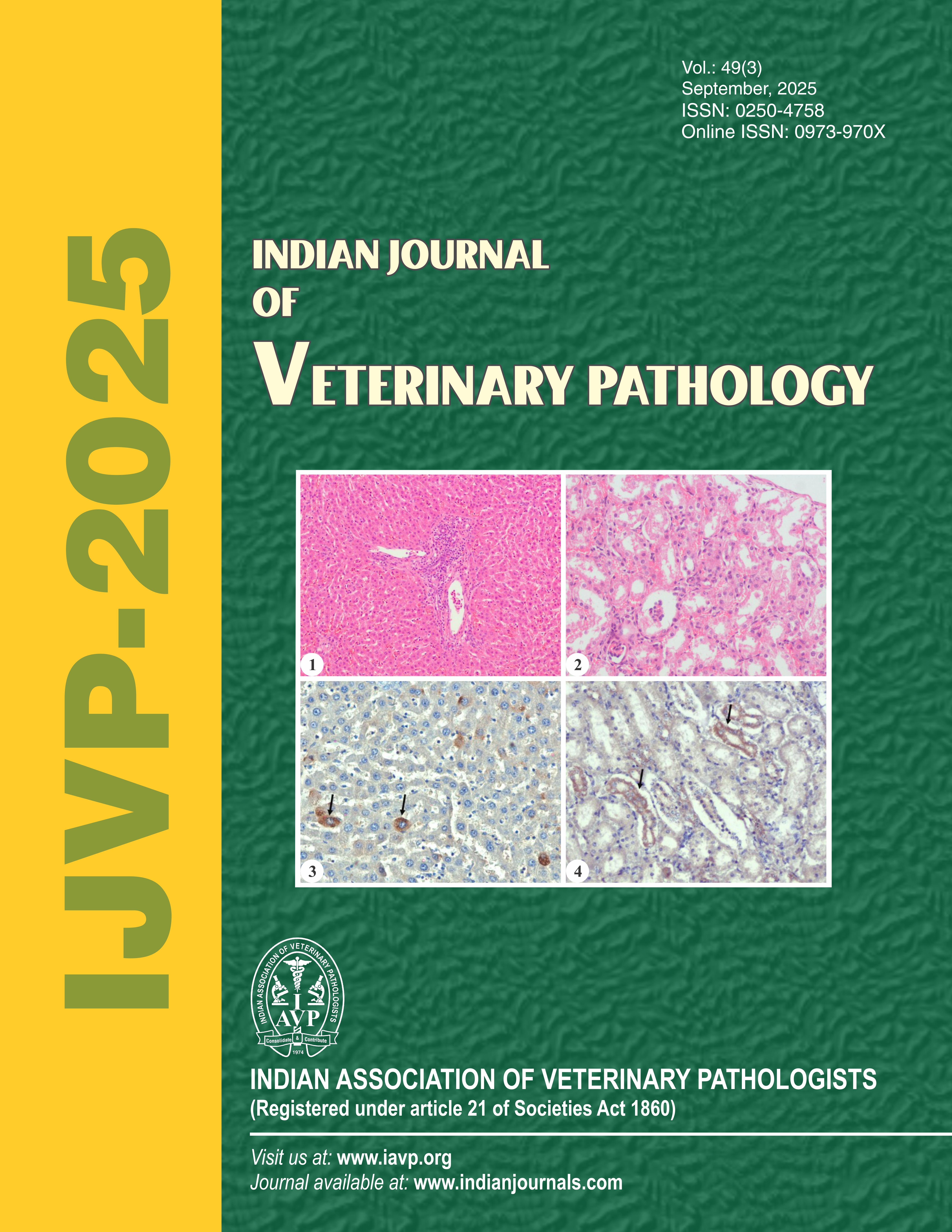
A widely metastatic hepatocellular carcinoma and intrahepatic bile duct cystadenocarcinoma in Spitz dog
184 / 146
Authors
-
 R. Madheswaran
Author
R. Madheswaran
Author
Keywords:
Dog, HCC, histopathology, ICC, immunohistochemistryAbstract
A ten-year-old male Spitz dog was brought for post mortem examination with a history of emaciation, weakness, difficulty in breathing and death. At necropsy, multiple tumour nodules were noticed on the liver surface. The metastatic tumour growths were also noticed in the lungs, lymph nodes and rectum. Morbid changes were observed in the spleen, heart, kidneys, urinary bladder and stomach. Microscopically, the tumour nodules were characterised by the presence of hepatocellular carcinoma (HCC) and intrahepatic bile duct cystadenocarcinoma (ICC). The pleomorphic neoplastic cells in HCC contained large, hyperchromatic, vesiculated nuclei, prominent nucleoli and multiple mitotic figures. The hepatic tissues around the tumour nodules were invaded with neoplastic cells and infiltration of mononuclear cells. The ICC consists of many cysts lined by single or multiple layers of cuboidal or columnar cells. The cystic lumen contained eosinophilic secretions and exfoliated cells. The metastatic neoplastic cells were noticed in the lungs, lymph nodes, spleen, heart, kidneys, urinary bladder, stomach and rectum. The immunoreactivity of neoplastic cells showed a mild expression of arginase-1, Hep Par-1, glypican-3, AFP and Bcl-2. The normal hepatic cells showed a marked expression of arginase-1, strong expression of Hep Par-1 and mild expressions of glypican-3 and AFP. Spleen showed marked expression of PCNA and CD3 in the lymphoid cells. Mesenteric lymph node showed a moderate expression of CD3 in the lymphoid cells.
Downloads
Downloads
Submitted
Published
Issue
Section
License
Copyright (c) 2025 Indian Journal of Veterinary Pathology

This work is licensed under a Creative Commons Attribution-NonCommercial-ShareAlike 4.0 International License.
How to Cite


