Cutaneous junctional melanocytoma in a dog: Clinical, cytological, histopathological and immunohistochemical evaluation
58 / 50
Authors
-
 S. Ramesh
Author
S. Ramesh
Author
Keywords:
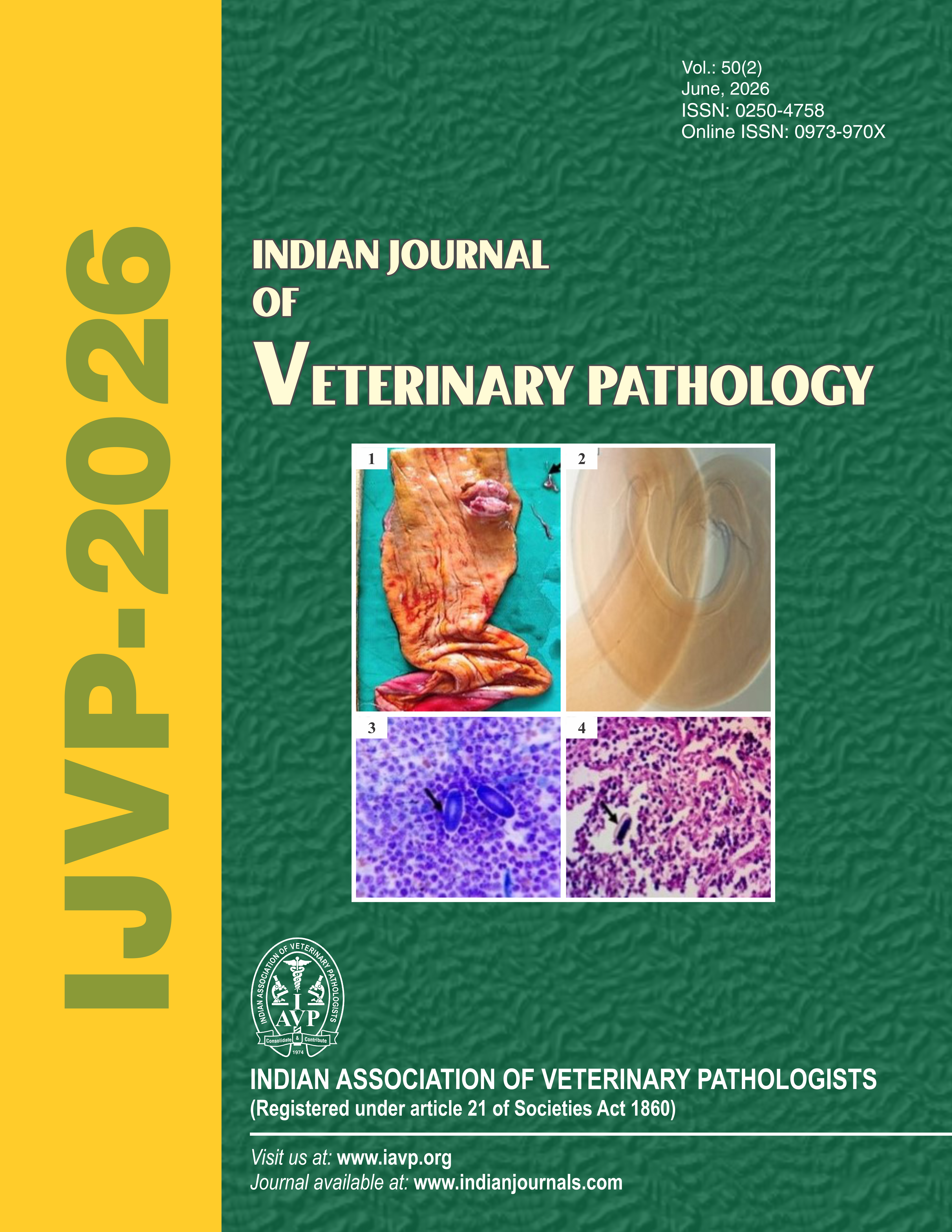
Cytology, dog, histopathology, immunohistochemistry, junctional melanocytomaAbstract
A 4-year-old Rottweiler dog had a black, firm nodule noticed in the dorsal aspect of a paw region in the periungual region at the nail bed/nail fold area of the paw. Cytology revealed brownish-black pigments dispersed throughout the cytoplasm and histopathology revealed melanocytic cells extending from the dermo-epidermal junction into the deep dermis with melanin granules. Immunohistochemistry confirmed strong Melan-A expression.
Downloads
Download data is not yet available.
Downloads
Submitted
2026-05-28
Published
2026-06-03
Issue
Section
Articles
License
Copyright (c) 2026 Indian Journal of Veterinary Pathology

This work is licensed under a Creative Commons Attribution-NonCommercial-ShareAlike 4.0 International License.
How to Cite
Cutaneous junctional melanocytoma in a dog: Clinical, cytological, histopathological and immunohistochemical evaluation. (2026). Indian Journal of Veterinary Pathology, 50(2). https://doi.org/10.56093/590j4k04


